Research Gives Hope to Stroke Survivors for Sight Recovery
May 2022
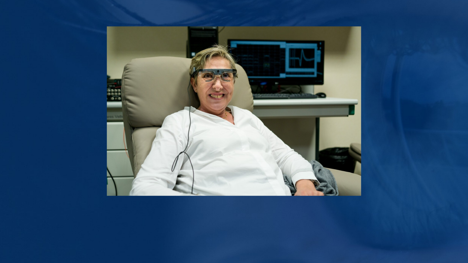
Researchers at the University of Nottingham, through the data obtained from clinical eye tests which consist of brain imaging to accurately map the areas of the brain that is affected by sight loss, have revealed insights that could allow identification of visual brain areas where functions could potentially be improved with rehabilitation.
In UK, around 150,000 people have a stroke and as a result, 30% of those experience some sort of a sight loss. Cerebral strokes are one of the common reasons which lead to visual field loss, which is referred to as hemianopia, where one side of a person’s sight or vision is affected and occurs due to the damage to the visual pathway to the brain.
In this new study, researchers combine perimetry, a visual field test, along with the multiple brain imaging datasets from four stroke survivors. This allowed them to see that the perimetry can be augmented with the brain imaging data to show a novel measurement of the residual visual field function. This potentially provides a personalized approach to therapy, guided by functional activity patterns in the post-stroke brain.
The research was led by PhD student Anthony Beh, under the supervision of Dr Denis Schluppeck and Prof Paul McGraw. Anthony explains, “A common misconception with stroke-related sight loss is that it affects vision through a particular eye. What is actually happening is that the eyes are seeing normally but the brain can’t process some of the information. This type of vision loss can be a particular problem for driving, reading or navigating a crowded space. It can also increase the risk of falls in older people. By exploring stroke-damaged brains with functional MRI and different kinds of visual stimulation, we found residual activity in the visual cortex, not detected by perimetry. This opens up possibilities for rehabilitation and offers new hope to stroke survivors.”
Dr Schluppeck adds: “By examining different types of brain scans we can actually see areas of ‘residual vision’ — places where the eyes and brain can still process images, even if this doesn’t reach awareness. Using MRI to pinpoint these areas of functional vision, clinicians could work with the stroke survivor and train them to recover some functionin that particular spot.”
This article was published by ScienceDaily.